Mammogram for Breast Cancer: How It Works, Accuracy, Benefits & What to Expect
Globally,breast cancer is the second most diagnosed cancer among women, following skin cancer. Less than 5% of all breast cancers in the U.S. are estimated to occur in women under age 40. Women age 45 and older have the greatest risk of developing breast cancer. The good news is that, over the past few decades, continuous developments in breast imaging and technology have made it possible to find many breast cancers even before symptoms appear. Mammography is considered the best screening technique and remains the most widely used method for breast cancer early detection.
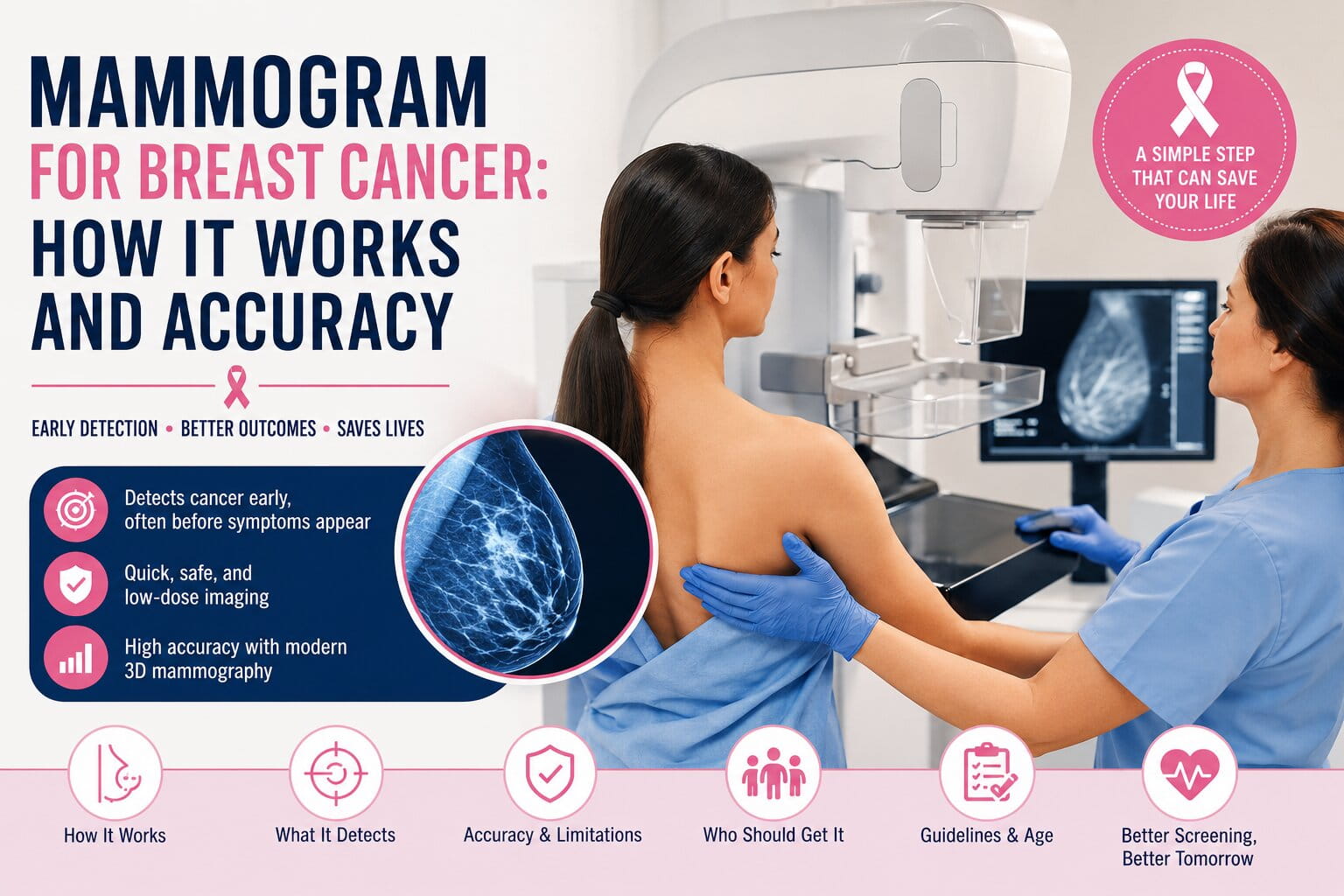
A mammogram can identify abnormalities such as tumors, microcalcifications, and suspicious tissue changes years before they become noticeable through a breast self-exam or clinical breast examination. Early detection often leads to more treatment options, less aggressive therapies, and better survival outcomes. While screening is essential, women should also understand the early signs of breast cancer and seek medical evaluation if they notice unusual changes between mammogram appointments.
This article addresses the mechanisms of mammography, the level of precision of the test, the study of what tumors can be visualized on a mammogram, and evaluating whether mammography is the best screening test for one’s self.

Mammogram for Breast Cancer: Key Takeaways
If you’re short on time, here are the most important facts about mammograms and breast cancer screening:
- A mammogram is the primary screening test used to detect breast cancer early.
- Mammograms can identify abnormalities years before symptoms develop.
- Screening mammography helps reduce breast cancer mortality through early detection.
- Mammogram accuracy generally ranges from 77% to 95%, depending on several factors.
- Dense breast tissue can make cancer harder to detect.
- 3D mammography may improve cancer detection and reduce false-positive results.
- Women at average risk should discuss mammogram screening beginning around age 40.
- Additional imaging tests such as breast ultrasound or MRI may be recommended in certain situations.
What Is a Mammogram?
A mammogram is a type of breast imaging test that makes use of very low levels of X-rays to check for cancer or other changes in the breast tissues.
Doctors and other healthcare professionals use mammography to:
- Conduct regular breast cancer screenings
- Discover new breast tumors
- Assess breast lumps
- Explore causes of nipple discharge
- Keep track of suspicious changes in breasts
- Follow up after breast cancer treatment
In fact, a mammogram is the primary screening method that doctors rely on to catch breast cancer at an early stage, even before symptoms appear.
Why Mammograms Are Important for Breast Cancer Detection
During the early phases, breast cancer grows silently. Most women diagnosed with early-stage breast cancer do not experience obvious breast cancer symptoms or warning signs at all, which is why routine screening plays such an important role in early detection.
Regular mammography assists in finding:
- Small breast tumors
- Ductal carcinoma in situ (DCIS)
- Invasive breast cancer
- Microcalcifications
- Structural tissue distortions
- Early-stage cancerous changesDuring the early phases, breast cancer grows silently. Most women diagnosed with early-stage breast cancer do not experience obvious breast cancer symptoms or warning signs at all, which is why routine screening plays such an important role in early detection.
Benefits of Early Detection
- Higher survival rates
- Earlier treatment intervention
- More treatment options
- Reduced risk of cancer spreading
- Better long-term outcomes
- Less extensive surgery in some cases
How Does a Mammogram Work?
Low-dose radiation is emitted through a mammogram to get highly detailed images of the internal breast tissue.
Step 1: Positioning
To accurately get breast images, a mammography technologist places a breast on the imaging platform.
Step 2: Compression
Then a compression paddle gently flattens the breast to get a thin layer.
Compression helps:
- Improve image clarity
- Spread tissue evenly
- Reduce motion blur
- Lower radiation exposure
- Detect smaller abnormalities
Step 3: Imaging
Multiple images are captured from different angles.
Step 4: Image Analysis
An expert radiologist examines the mammogram and searches for:
- Breast masses
- Calcifications
- Tissue asymmetry
- Distortions
- Suspicious lesions
Most mammogram appointments take between 15 and 30 minutes.
Types of Mammograms
Screening Mammogram
A screening mammogram is performed on women who show no signs or symptoms of breast cancer.
Its main aim is to detect cancer at a stage when there are still no symptoms.
Common Uses
- Annual breast cancer screening
- Preventive healthcare
- Routine breast monitoring
Diagnostic Mammogram
A diagnostic mammogram is performed when symptoms or abnormalities are present.
Common reasons include:
- Breast lump
- Nipple discharge
- Breast pain
- Abnormal screening results
- Skin changes affecting the breast
Diagnostic mammograms typically involve additional images and closer evaluation.
Digital Mammography
Digital mammography stores images electronically rather than on traditional film.
Benefits include:
- Better image storage
- Easier image comparison
- Faster image review
- Improved workflow
3D Mammography (Breast Tomosynthesis)
3D mammography creates multiple image slices of breast tissue that can be viewed layer by layer.
Advantages
- Improved cancer detection
- Better evaluation of dense breast tissue
- Fewer false positives
- Reduced recall rates
Many breast imaging centers now prefer 3D mammography for routine screening.
What Can a Mammogram Detect?
Mammograms can identify abnormalities such as tumors, calcifications, and early-stage cancers before they become noticeable. Detecting cancer at an earlier stage often improves treatment outcomes and survival rates.
Mammograms Can Detect:
- Breast cancer
- Breast tumors
- Microcalcifications
- Tissue distortions
- Benign breast masses
- Fibroadenomas
- Certain breast cysts
- Ductal carcinoma in situ (DCIS)
Early detection of these abnormalities allows healthcare providers to recommend further testing as part of the breast cancer diagnosis process and begin treatment when necessary.
How Accurate Is a Mammogram?
Mammography is a highly effective tool; however, no screening test is 100% perfect.
A number of things can affect the accuracy, such as the density of the breast, age, tumor location, and imaging technology.
| Accuracy Measure | Typical Range |
| Sensitivity | 77–95% |
| Specificity | 90–95% |
| Cancer Detection Rate | High |
| False Positive Rate | Moderate |
In general, mammograms correctly identify most breast cancers while minimizing unnecessary procedures.
Understanding Mammogram Sensitivity and Specificity
Ultrasound imaging uses the very short wavelength sound waves that travel through the body tissues and locate the target organs and abnormal tissues inside.
Sensitivity
It’s the term that defines how well a mammogram detects cancer cases, actually.
An increase in sensitivity results in a decrease in the number of undetected cancer cases.
Factors That Improve Sensitivity
- 3D mammography
- Less dense breast tissue
- High-quality imaging
- Experienced radiologists
Specificity
Specificity is defined as the ability to correctly exclude the presence of cancer in healthy women.
Improvement in specificity can significantly lower:
- Unnecessary biopsies
- Anxiety
- Additional testing
Factors That Affect Mammogram Accuracy
Dense Breast Tissue
Dense breast tissue is one of the most important factors affecting mammogram performance.
Because both dense tissue and tumors appear white on mammograms, cancer may be harder to detect.
Women with dense breasts may benefit from:
- Breast ultrasound
- Breast MRI
- 3D mammography
Age
Mammograms generally become more accurate with age because breast tissue often becomes less dense.
Tumor Characteristics
Certain cancers are more difficult to visualize, including:
- Lobular carcinoma
- Fast-growing tumors
- Deep tissue lesions
Imaging Technology
Newer technologies, particularly 3D mammography, may improve cancer detection rates.
Dense Breasts and Breast Cancer Risk
Dense breast tissue is common and affects millions of women.
Breasts are considered dense when they contain more glandular and fibrous tissue than fatty tissue.
Why Dense Breasts Matter
Dense breast tissue can:
- Make mammogram interpretation more difficult
- Increase the chance of a missed cancer
- Slightly increase breast cancer risk
Women with dense breasts should discuss supplemental screening options with their healthcare provider.
Additional imaging may include:
- Breast ultrasound
- Breast MRI
- 3D mammography
Can Mammograms Miss Breast Cancer?
Yes. This is called a false-negative result.
Reasons may include:
- Dense breast tissue
- Small tumors
- Hidden lesion locations
- Aggressive cancer growth
Women should always discuss concerning symptoms with their healthcare provider, even if a mammogram result is normal.
Can Mammograms Produce False Positives?
Yes.
A false positive occurs when mammography suggests cancer but follow-up testing confirms no cancer is present.
This may result in:
- Additional mammograms
- Breast ultrasound
- MRI testing
- Breast biopsy
Although stressful, false positives help ensure suspicious findings receive proper evaluation.
Mammogram Results Explained: Understanding BI-RADS Scores
Radiologists commonly use the Breast Imaging Reporting and Data System (BI-RADS) to classify mammogram findings.
Common BI-RADS Categories
- BI-RADS 0: Additional imaging needed
- BI-RADS 1: Negative result
- BI-RADS 2: Benign finding
- BI-RADS 3: Probably benign
- BI-RADS 4: Suspicious abnormality
- BI-RADS 5: Highly suggestive of cancer
- BI-RADS 6: Known cancer diagnosis
Understanding your BI-RADS score can help clarify whether further testing is needed.
What Happens If Your Mammogram Is Abnormal?
Receiving an abnormal mammogram result can be frightening, but it does not automatically mean breast cancer is present.
Many abnormal findings turn out to be benign (noncancerous).
Possible next steps may include:
- Additional mammogram views
- Diagnostic mammography
- Breast ultrasound
- Breast MRI
- Breast biopsy
Common noncancerous findings include:
- Breast cysts
- Fibroadenomas
- Benign calcifications
- Dense breast tissue changes
Your healthcare provider will explain the findings and determine whether additional testing is necessary.
Understanding Breast Biopsies After a Mammogram
If an abnormality appears suspicious, a breast biopsy may be recommended.
A biopsy involves removing a small sample of breast tissue for laboratory analysis.
Types of breast biopsies include:
Fine Needle Aspiration
Uses a thin needle to collect fluid or cells.
Core Needle Biopsy
Removes small tissue samples using a larger needle.
Surgical Biopsy
Removes part or all of a suspicious area for evaluation.
A biopsy is the only way to definitively determine whether breast cancer is present.
Signs You May Need a Diagnostic Mammogram
Contact your healthcare provider if you notice:
- A breast lump
- Nipple discharge
- Breast skin dimpling
- Nipple inversion
- Persistent breast pain
- Breast swelling
- Enlarged lymph nodes under the arm
These symptoms do not automatically mean cancer is present, but they warrant medical evaluation.
Mammogram vs Ultrasound
| Feature | Mammogram | Ultrasound |
| Routine Screening | Yes | No |
| Detects Calcifications | Excellent | Limited |
| Dense Breast Evaluation | Moderate | Better |
| Radiation | Yes | No |
Ultrasound is often used as a supplemental imaging tool rather than a replacement for mammography.
Mammogram vs MRI
MRI is generally reserved for women at high risk of breast cancer.
MRI May Be Recommended For:
- BRCA1 mutations
- BRCA2 mutations
- Strong family history
- Previous chest radiation exposure
- High-risk screening programs
Is 3D Mammography Worth It?
Many women wonder whether upgrading to 3D mammography is worth the additional cost or effort.
For many patients, especially those with dense breast tissue, the answer may be yes.
3D mammography (breast tomosynthesis) creates multiple image slices of the breast rather than a single flat image. This allows radiologists to examine breast tissue layer by layer.
Benefits of 3D Mammography
- Improved breast cancer detection rates
- Better visualization of dense breast tissue
- Fewer false-positive findings
- Reduced need for repeat imaging
- Increased diagnostic confidence
Potential Limitations
- Slightly higher cost in some locations
- Not available at all imaging centers
- Insurance coverage may vary
For women with dense breasts or previous abnormal mammograms, 3D mammography may provide significant advantages over traditional 2D screening.
Who Should Get a Mammogram?
Women at average risk should discuss screening with their healthcare provider beginning around age 40.
Higher-risk women may require earlier screening.
Risk Factors Include
- Family history of breast cancer
- BRCA gene mutations
- Previous breast cancer diagnosis
- Dense breast tissue
- Prior radiation exposure
Mammogram Screening Guidelines by Age
Breast cancer screening recommendations may vary slightly among medical organizations, but most experts agree that routine mammography plays an important role in preventive healthcare.
Ages 40–49
Women should discuss breast cancer risk factors and screening options with their healthcare provider.
Benefits of screening may include:
- Earlier cancer detection
- Reduced risk of advanced-stage disease
- Increased treatment options
Ages 50–74
Routine mammography is generally recommended for most women in this age group.
Many healthcare organizations recommend screening every one to two years.
Age 75 and Older
Screening decisions should be based on:
- Overall health
- Life expectancy
- Personal preferences
- Physician recommendations
Women who remain in good health may continue benefiting from routine mammography.
Risk Factors That Increase the Need for Mammography
While all women should understand breast cancer screening recommendations, certain individuals may have a higher-than-average risk.
Family History of Breast Cancer
Risk increases when close relatives have been diagnosed with breast cancer.
Examples include:
- Mother
- Sister
- Daughter
BRCA1 and BRCA2 Gene Mutations
Inherited genetic mutations can significantly increase lifetime breast cancer risk.
Women with these mutations often begin screening earlier and may undergo MRI screening in addition to mammography.
Previous Breast Cancer
Women previously treated for breast cancer require ongoing surveillance imaging.
Previous Chest Radiation
Radiation therapy involving the chest area may increase future breast cancer risk.
How to Prepare for a Mammogram
Preparing properly can help improve image quality and make the experience more comfortable.
Before Your Appointment
- Schedule the exam when breasts are less tender if possible.
- Wear a two-piece outfit.
- Bring previous mammogram records.
- Inform staff of any breast symptoms.
Avoid Using
- Deodorant
- Powder
- Lotion
- Perfume
These products can sometimes appear on mammogram images and affect interpretation.
Mammogram Cost and Insurance Coverage
The cost of a mammogram varies depending on:
- Location
- Imaging center
- Type of mammogram
- Insurance coverage
Many health insurance plans cover routine preventive mammography according to screening guidelines.
Before scheduling an appointment, verify coverage details with your insurance provider.
Can a mammogram detect all breast cancers?
No. While mammograms are highly effective, some cancers may not be visible, especially in women with dense breast tissue.
How often should I get a mammogram?
Screening frequency depends on age, risk factors, and healthcare provider recommendations. Many women undergo mammography every one to two years.
What happens if my mammogram shows something suspicious?
Additional testing such as diagnostic mammography, ultrasound, MRI, or biopsy may be recommended.
Are mammograms safe?
Yes. Mammograms use very low-dose radiation and are considered safe for routine screening.
Can younger women get mammograms?
Yes. Women with higher breast cancer risk factors may begin screening earlier than average-risk women.
Are mammograms covered by insurance?
Many health insurance plans cover routine breast cancer screening mammograms, though coverage varies by provider and location.
Can men get mammograms?
Although breast cancer is less common in men, mammography may sometimes be used when suspicious breast symptoms are present.
Is mammography effective for dense breasts?
Mammography remains valuable, but supplemental imaging tests may improve detection in women with dense breast tissue.
Wrapping Up
Mammography remains one of the most effective tools available for detecting breast cancer early. By identifying abnormalities before symptoms develop, mammograms can improve treatment outcomes, expand therapeutic options, and save lives.
Whether you’re preparing for your first mammogram, comparing screening options, or trying to understand your results, staying informed about breast cancer screening is an important step toward protecting your long-term health.
Women should discuss their personal risk factors, family history, breast density, and screening preferences with their healthcare provider to determine the most appropriate breast cancer screening strategy.
- American Cancer Society: Breast Cancer Screening Guidelines
- National Cancer Institute: Mammograms and Breast Cancer Screening
- CDC: Breast Cancer Screening
- RSNA: Mammography Information for Patients
- ACR: Breast Imaging and Screening Recommendations
- USPSTF: Breast Cancer Screening Recommendations
- WHO: Breast Cancer Facts and Early Detection Resources
- Peer-reviewed studies published in journals such as:
JAMA (Journal of the American Medical Association)
The New England Journal of Medicine (NEJM)
Radiology
The Lancet Oncology
Breast Cancer Research and Treatment


A mammogram is a low-dose X-ray of the breast used to detect breast cancer and other abnormalities. Modern mammography can detect approximately 77%–95% of breast cancers, depending on factors such as breast density, age, tumor characteristics, and imaging technology used. 3D mammography (tomosynthesis) often improves detection rates and reduces false-positive results compared with traditional 2D mammograms.