Breast Cancer Diagnosis: Tests, Procedures & Detection Methods Explained
Breast cancer diagnosis is a structured clinical evaluation process used in oncology and radiology to detect, confirm, and stage malignant breast tissue changes related to Breast Cancer: Causes, Symptoms, Stages, Treatment & Prevention.It involves a combination of screening methods, diagnostic imaging, and tissue examination via microscopy to allow the accurate identification of both early and late stages of breast cancer.
Contemporary diagnostic strategies are founded on medical guidelines that have been validated by research and involve a combination of radiological imaging and pathology to minimize the occurrence of false results and lessen the number of patients undergoing unnecessary surgeries.
Besides, early and precise diagnosis helps a lot in reducing breast cancer fatalities and adverse side effects of treatments by using chemotherapy at the right time, surgery, and other techniques.
What Is Breast Cancer Diagnosis?
Breast cancer diagnosis involves a series of clinical and laboratory steps aimed at identifying cancerous tumors in the breast. The process entails a team effort among radiologists, oncologists, and pathologists to not only diagnose the presence of the disease but also to understand the biological characteristics of the tumor.
Women experiencing suspicious symptoms should understand the early signs of breast cancer for earlier clinical evaluation.
Clinical Objectives:
- Detecting cancerous breast lesions at an early stage
- Obtaining a histopathological basis for the presence of cancer cells
- Categorizing the tumor (ductal, lobular, invasive, etc.)
- Staging based on metastasis and lymph node involvement
- Development of personalized treatment strategy
Clinical Symptoms That Trigger Diagnostic Evaluation
Diagnostic testing usually starts when patients with suspicious clinical findings or abnormal screening results are referred to a doctor for further evaluation.
Common Clinical Signs:
- Discovery of a breast or axillary lump by touch (lump)
- Breast asymmetry or structural changes
- Skin dimpling or the appearance of orange peel
- Nipple inversion or a discharge that is bloody or serous
- Persistent localized breast pain or tenderness
- Inflammatory signs – skin redness or swelling
These signs should be the focus of clinical assessment without delay for malignancy exclusion.
Step-by-Step Diagnostic Workflow
Diagnosis of breast cancer involves following a clinical pathway that has been standardized, which includes check-up of the physical condition, different ways of getting pictures for imaging diagnostics, and tissue biopsy.
1. Clinical Breast Examination (CBE)
This examination is done by a medical professional to find out if there are any abnormalities by touch in the breast and axillary lymph nodes.
Clinical Purpose:
- To estimate the risk for the first time
- Finding lumps that can be felt
- Checking lymph nodes
A non-invasive and simple assessment tool first used in oncology screening protocols
2. Diagnostic Imaging Techniques
Imaging using radiology is very useful in breast cancer because it shows the structure of the breast tissue and help the doctors to identify the abnormal ones.
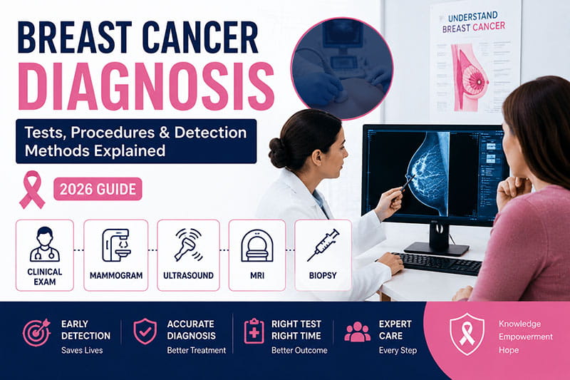
Mammography (Primary Screening Modality)
Mammography is a very reliable low-dose X-ray imaging tool that is used for early detection of breast cancer, especially in asymptomatic patients.
Clinical Advantages:
- Spotting microcalcifications that occur in association with ductal carcinoma in situ (DCIS)
- Finding tumors that are still in the early stages and have not yet manifested symptoms
- The most widely used screening method for women 40 years and older
Limitations:
- Very dense breast tissue can mask tumors, leading to false negatives
- Additional imaging, such as MRI, tomography, or ultrasound, can be necessary for certain breast cancers
Breast Ultrasound (Sonographic Evaluation)
Ultrasound imaging uses the very short wavelength sound waves that travel through the body tissues and locate the target organs and abnormal tissues inside.
Clinical Use Cases:
- Locating a breast lump that can be felt through the skin
- Assessment in younger patients with dense breast tissue
- Guidance for needle biopsy procedures
Breast MRI (Magnetic Resonance Imaging)
MRI is capable of producing high-resolution images of soft tissues with the help of magnetic fields and contrast agents.
Indications:
- Genetically high-risk individuals (e.g., carriers of BRCA1/BRCA2 mutations)
- Planning surgery for tumor removal
- Uncovering multifocal or hidden lesions
3. Tissue Biopsy (Definitive Histopathology)
Biopsy is the final, most reliable procedure to determine if the tumor is really cancerous.
Histopathological Process:
A tissue sample is extracted and analyzed under a microscope to detect cancerous cellular morphology.
Types of Biopsy:
- Fine Needle Aspiration (FNA)
- Core Needle Biopsy
- Excisional (Surgical) Biopsy
Diagnostic Output:
- Tumor grade (aggressiveness)
- Receptor status (ER, PR, HER2)
- Histological classification
Diagnostic Decision Pathway (Clinical Reasoning)
Diagnosing breast cancer involves a recognized series of steps that are followed very closely:
- Mammogram screening for a possible first detection
- Ultrasound/MRI for checking the features of the lesion
- Biopsy for histology confirmation
- Pathology report for final diagnosis
Stepwise approach like this one drastically reduces diagnostic errors and helps to ensure clinical accuracy.
Types of Breast Cancer Identified Through Diagnosis
Histopathological analysis reveals the nature of different cancer types:
- Invasive Ductal Carcinoma (IDC)
- Invasive Lobular Carcinoma (ILC)
- Ductal Carcinoma In Situ (DCIS)
- Triple-Negative Breast Cancer (TNBC)
Different cancer subtypes have different growth features, drug susceptibility, and treatment patterns.
Staging and Disease Progression Analysis
The TNM system is applied by an oncologist to determine the stage, after the diagnosis, which is TNM staging (Tumor, Node, Metastasis).
Clinical Stages:
- Stage 0: In situ carcinoma (non-invasive)
- Stage I–II: Localized disease
- Stage III: Regional lymph node involvement
- Stage IV: Metastatic systemic spread
Diagnostic Accuracy and Clinical Reliability
Sensitivity & Specificity Overview:
| Test | Diagnostic Role | Reliability |
| Mammography | Screening | High |
| Ultrasound | Characterization | Moderate |
| MRI | High-risk detection | Very High |
| Biopsy | Definitive diagnosis | 100% (Gold Standard) |
Combined diagnostic modalities significantly reduce false-negative rates.
Importance of Early Clinical Detection
Diagnosis at an early stage will directly improve oncological outcomes by allowing less aggressive treatment regimens. Healthy lifestyle habits and lifestyle changes that may reduce breast cancer risk may further improve outcomes.
Clinical Benefits:
- Elevated 5-year survival rates
- Lowered chemotherapy dependency
- Better breast conservation effectiveness
- Decreased risk of metastasis
Nutritional support through a proper breast cancer diet plan and regular physical activity may also support recovery.
When to Seek Medical Evaluation
It is highly advised to get a clinical assessment without delay when:
- A new lump in the breast is discovered
- There are changes in the skin or nipple
- The breast pain or discomfort is ongoing
- The results from screening are not normal
Emotional stress during diagnosis may affect mental health during cancer treatment and overall family well-being.
What is the most accurate test for breast cancer?
Although imaging techniques may show suspicion, it is the Histopathological biopsy that ultimately determines the presence of cancer.
Can imaging alone confirm breast cancer?
No, imaging suggests suspicion but biopsy confirms malignancy.
What is the role of mammography in diagnosis?
It serves as the primary screening tool for early detection. alongside the latest advances in latest breast cancer treatments.
Is breast cancer diagnosis painful?
Most imaging tests are non-invasive; biopsy may involve mild discomfort.
How is cancer stage determined?
Through TNM staging based on tumor size, lymph node involvement, and metastasis.
Wrapping Up
Initially, diagnostic mammography and high-frequency ultrasound can be used to find suspicious tumors. Breast cancer diagnosis is an interdisciplinary clinical procedure that includes radiology, oncology, and pathology. The combination of mammography, ultrasound, MRI, and biopsy allows for quite accurate detection and staging of malignant breast tumors.
Public education, breast cancer awareness and early detection, and supportive recovery strategies remain essential for improving long-term outcomes. Some patients also explore natural remedies to support breast cancer recovery alongside conventional medical treatment and lifestyle modifications.
The ability to diagnose accurately and early is the most vital factor for success in treatment, patient effectiveness, and overall survival rates.
- World Health Organization (WHO): Breast Cancer Facts
- American Cancer Society: Breast Cancer Information
- National Cancer Institute: Breast Cancer Diagnosis & Treatment
- Mayo Clinic: Breast Cancer Symptoms & Diagnosis
- Breastcancer.org: Screening & Diagnostic Tests
Early signs of breast cancer can include a new lump in the breast or underarm, changes in breast size or shape, and unusual nipple discharge. You may also notice skin dimpling, redness, or nipple changes that should never be ignored. These symptoms are often subtle at first, but early detection is crucial for effective treatment and better survival outcomes.